
The pre-operative eye examination is the key for a successful surgical procedure. It is the most important source of information for your eye doctor, who will evaluate the state of your eyes and choose the best suited surgical method for you. The same goes for your eye surgeon, who will be operating on you. That is the reason why the quality and extent of data obtained during the preoperative examination is so important.
At our clinics in Bratislava, Brno, Český Těšín and Prague we know that each and every one of you has individual expectations from their vision, different hobbies, varying types of jobs. Additionally, every single eye is different. That is why we give each and every one of our patients individual care and attention.
We are constantly investing into new machine technology and equipment, so that we can always offer the highest possible level of health care.
We reach high- quality results of the examinations through a synergy of the work of the whole team. That is the reason why our mutual collaboration and the Czech and Slovak consillia are important to us, as well as our collaboration with you, our patients. Please follow the instructions of our doctors, nurses and optometrists during the examinations.

The procedure is the following: starting with basic data concerning the visual acuity and intraocular pressure, followed by parameters of the anterior eye segment (the cornea, the iris and the eye lens), a test of the actual extent of the refractive error, leading up to the detailed description and exploration of the so-called posterior eye segment (the retina, the vitreous body).
The importance of dilating the eyes
In order to examine the retina and explore the actual extent of the refractive error, it is necessary to dilate the eye with the help of eye drops. It is necessary to put the eyes into a state, where the muscles responsible for focusing (the so- calles cycloplegia) are „paralyzed“. This examination is crucial for the doctor to be able to decide whether you are a suitable candidate for the laser operation or not.
A myopic eye (most of the patients who are doing a laser surgery have to undergo this because of myopia) is „longer“ than a healthy eye. Additionally, it suffers from serious retinal diseases more often. The retina is often thinner, and the changes usually also affect the retina's edges (the retinal peripheries). If the doctor were to not notice any retinal disorders or risky changes in its peripheries before a laser surgery or any other eye surgery, the consequences could be fatal – a retinal detachment or even a complete loss of vision. A high- quality and detailed examination of the retina cannot be done without dilating the retina first.
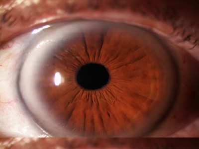
The retina without dilation
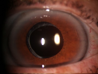
When the retina is dilated, the doctor can very accurately see the whole retinal surface, including its peripheral areas. That way, any visible or hidden degeneration can be uncovered. If the patient is undergoing the entry examination and the laser surgery on the same day, it is clear that he could not have undergone the examination with a dilated retina. A patient, who has had their retina dilated, cannot undergo the most modern laser surgery on the same day,because the laser monitor (the eye tracker) that is responsible for the accuracy of the procedure, would not work.
-The refractive error will not be removed from the eye to the full extent. The procedure will have to be repeated. Because of this, you should always choose a clinic that offers additional corrections (additional laser treatment - a second surgery) within the basic price of the surgery. The correctional surgery could raise the price of the original surgery to twice the amount!
- More dioptries could be removed than necessary – which could lead to an exactly reversed refractive error. In this case, an additional correction would be even more complicated, and you could be left with reading glasses or glasses for long distances.
- A previously uncovered retinal disorder could arise – a tear, retinal detachment, bleeding into the vitreous body and other problems, which can cause a worsening or complete loss of vision.
The eye is one of our most sensitive organs, so you should consider each and every detail when choosing an eye clinic. One of the most important ones is the quality and precision of the entry examination.
„Before a laser or intraocular surgery, it is inevitable to explore the full extent of the entry examination. This is the only way for us to be able to recommend the best solution to our patient, taking into account their age and predicting the most probable future development of the eye. One part of a complex entry examination is definitely the retinal examination during mydriasis, or the so- called dilation. This examination should be performed by a doctor, who has experience with the diagnostics of the posterior eye segment and retinal disorders. The absence of this dilation could actually put the patient at risk,“ our doctors and eye surgeons agree.
- patient history (the exploration of your medical and ocular history),
- an examination of your visual acuity and measurement of your intraocular pressure
- an examination of the thickness, curvature and diameter of the cornea
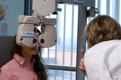
- an examination of your binocular vision (the collaboration of both eyes) and the subjective refraction by an optometrist with the help of trial glasses, or the more modern method, with the help of a computer phoropter
- an examination of the surroundings of the eye and the anterior eye segment (the cornea, the anterior chamber, the iris, the eye lens)
- an examination of the ocular background (the retina and the vitreous body, the optical nerve – the place of the sharpest vision) by a doctor
The patient history is performed by a professionally trained nurse and a doctor. The goal is to gather the following information:
• the character of the patient´s problems
• information about the current and past development of the eye, vision and optical error
• information about previous eye surgeries, injuries and treatments
• general diseases and medication that could affect the health condition of the eye and its individual tissues or the optical error and the postoperative development
• since many of these diseases have a genetic relation, it is very beneficial for the doctor to have all the available information about general and optical diseases in the immediate family of the patient
the autorefractometer- measures the objective refractive error of the eye
keratometer - measures the corneal curvature of the eye
a contactless, or optionally applanation tonometer - measures the intraocular pressure
pentacam - a modern imaging device used for the examination of the anterior segment of the eye. Based on the obtained images, a number of parameters can be described accurately and in detail, including the map of the corneal thickness, the anterior chamber, the topography of the cornea, clarity and eventual cataracts of the eye lens etc.
aberometer - measures the irregularity of the whole optical system of the eye, including different deviations. It is mostly used for errors which are difficult to describe and cannot be corrected with the help of the usual corrective devices (dioptric glasses, contact lenses). They can, however, significantly lower the quality of vision, especially when combined with a wide pupil (young patients, genetic predispositions, in twilight...).
Following these examinations, the patient will be examined by an optometrist, who is best suited to explore the dioptric correction and the visual acuity of both eyes with the help of a modern machine, the phoropter. He measures the ability of depth perception, the collaboration of both eyes, the optical dominance, the balance of the muscles responsible for moving the eyes, and eventually also the contrast sensitivity.
• loosening of the accommodative muscles (the so- called cycloplegia). Thanks to the cycloplegia, it is possible to discover hidden dioptries. People who are born with presbyopia have the ability to compensate for their dioptries with constant focusing. On the other hand, in patients with myopia, this error can, in some rare cases, be caused by the long- term flexing of the focusing mucles, even if they do not suffer from a refractive error. The cause can also be neurological or lie in hidden strabism. If this step is underrated, the dioptries can either „stay put“ after the laser or intraocular surgery, or new ones can develop. The patient could have even more problems after the surgery than before. After a certain time has passed, the patient would need to undergo the surgery again, if it is even possible in such a case. The detailed examination of the state of the retina is performed by a professional eye doctor and it needs to be given the appropriate amount of attention. Changes in the retina that are caused by hidden diseases usually cannot be corrected and cause a worsening vision, blackouts in the field of vision or even a complete loss of vision.
After the retinal examination and the examination of the posterior segment of the eye and a conversation with the patient, the doctor will be able to recommend the most suitable method of treatment for the specific refractive error, or eventually other treatment methods.
The whole process takes approximately 2 – 3 hours and results in perfect materials that a successful surgical procedure can be based on.
You will be able to undergo the surgery during the next few days, but not on the same day as the entry examination. This small hold- up is very beneficial for the safety of your eyes. This is a time when hurrying would be absolutely useless. In the case of the entry examination, we should „hurry slowly“, so we do not have to regret it the next day.